Hone your POCUS interpretation and clinical reasoning by working through the interactive case below.
Case #15: Not the Most Phlegm-buoyant
Ms C is a 95-year-old female with a past medical history of hypertension and CKD. She presented with hypoxia, orthopnea, lower limb edema, and chest discomfort for 2 days.
CXR reported left lower lobe patchy consolidation and prominence of interstitial markings. BNP was 40,000. She was admitted for pneumonia and CHF. In addition, troponin on admission was 1854 → 1848 → 1180. TTE done showed reduced EF 30%. Cardiology impression was ?missed MI vs stress cardiomyopathy. She was started on DAPT, PO cefuroxime, and diuretics. She received IV furosemide 40mg qdaily for 3 days and transitioned to PO furosemide 40mg qdaily for past 2 days.
POCUS was consulted for ongoing dyspnea and hypoxia requiring 3L of oxygen.
Lung Scans
Right anterior lung field
Right lateral lung field
Left anterior lung field
Left lateral lung field
Right PLAPS
Left PLAPS
Cardiac Scans
Parasternal long axis
Apical 4-chamber
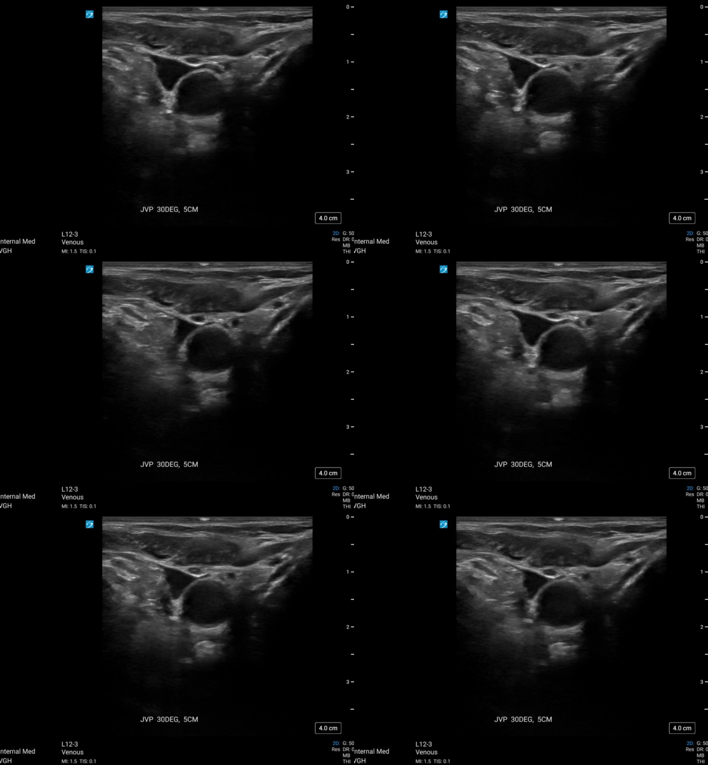
JVP and IVC
Case continued
Given her clinical deterioration with hypoxemia/dyspnea, POCUS recommendations were as follows:
1. Escalate antibiotic coverage to IV (such as ceftriaxone/azithromycin)
2. CT chest for further characterization of lung parenchyma
3. Increase furosemide to 40 mg IV daily
Later in the week, the POCUS team was asked to reassess the patient in the context of increased work of breathing (respiratory rate 30) and worsening hypoxemia (requiring 7 L oxygen via facemask).
Lung scans
Anterior lung fields
Right lateral lung field
Right PLAPS
Left lateral lung field
Left PLAPS
IVC
Case continued
CT chest showed “Extensive asymmetric ground-glass opacities throughout all lobes most significant involvement of the upper lobes. There is associated interlobular septal thickening and bronchial wall thickening. Appearance is most suggestive of a combination of pulmonary edema and atypical infection.” Overall consistent with our ultrasound findings.
Case continued
A few days later, she demonstrated significant improvement. Oxygen requirements were down to 2L and she was much more comfortable with normal work of breathing.
NT-pro BNP was done and >35000. Because of this, we were asked to assess for pulmonary edema to guide further diuretic therapy.
Scans at 2nd Follow up
IVC
Right anterior lung field
Right lateral lung field
Left anterior lung field
Left lateral lung field
Case Conclusion
POCUS again showed an interstitial lung syndrome with irregular B-line pattern mainly in the upper lung zones and ragged pleura. Overall suggesting an infection/inflammatory cause for her hypoxemia. CVP no longer elevated.
She was weaned off oxygen, and converted from IV to PO furosemide and was eventually put back on her home dose. BNP remained high though trending down from >35 000 to 25 000. The elevated BNP was attributed to the acute insult and ?Takotsubo cardiomyopathy vs missed ACS.


